In this article
View 4 More +Finding any type of growth on your beloved companion can certainly be concerning, but it is important to keep in mind that not all growths are cancerous, and some may not cause any problems for your pet. Cysts are a type of growth that dogs of any age and breed may experience. They are hollowed-out structures formed within tissues, and these structures can fill with dead cellular material or secretions made by the body.
It is important to note that cysts can occur internally in the body as well, impacting some organs, including the kidneys, ovaries, and spleen. In this article, we will explore common cysts impacting the skin of dogs, which typically are benign. Continue reading to learn about the five most common cysts impacting the skin of dogs.

The 5 Types of Cysts on Dogs
1. True Cysts
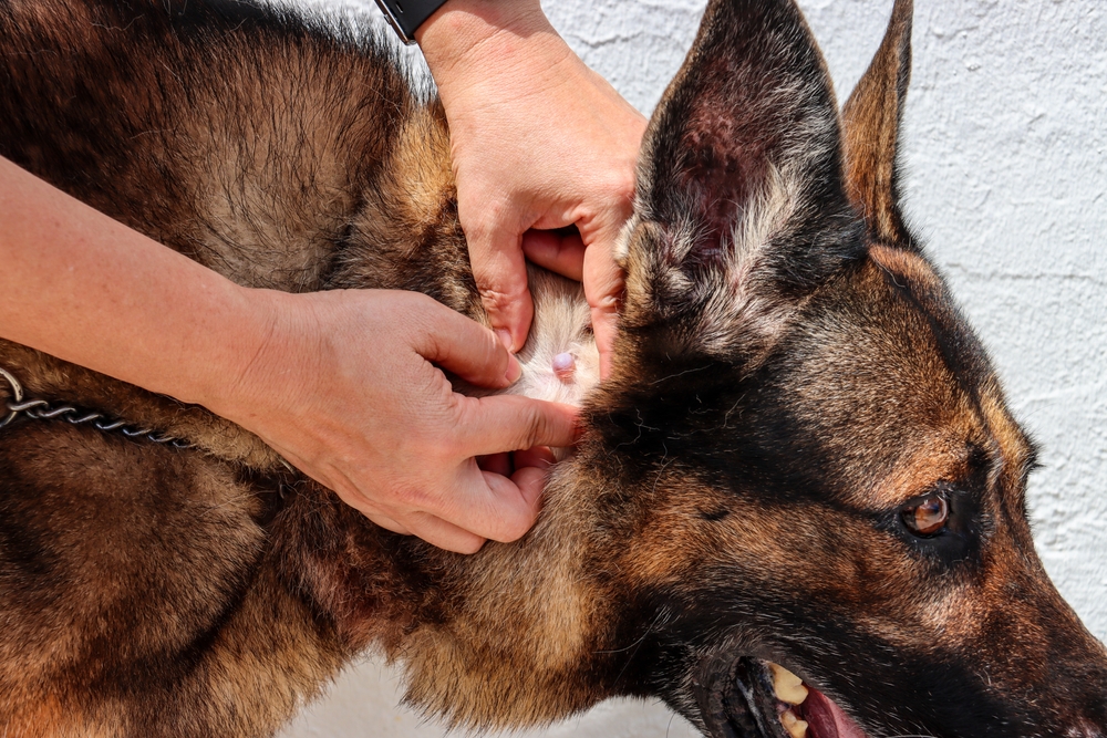
True cysts typically form within glands, particularly sweat glands, as the result of a blocked duct. True cysts have a lining within the structure that produces secretions. True cysts are common in both dogs and cats, and cysts can appear clear and resemble a vesicle. These cysts often contain a watery fluid and tend to occur on the neck, head, and back. Even if your dog has a cyst that looks to you like a true cyst, it’s best to get them checked out by the vet, as they may appear similar to other skin changes or may be mistaken for more serious cysts or lumps.
2. Follicular Cyst
Follicular cysts occur within hair follicles and cause a dilation of the follicle. Follicular cysts can occur anywhere on the body; however, pressure points and areas with friction may be more prone. These types of cysts typically contain fluid or dark, thick secretions that can contain keratin.
A common type of follicular cyst is an interdigital cyst, which occurs between toes. Interdigital cysts are commonly correlated to allergies, and treating allergies can benefit the dog’s skin health and reduce the level of itchiness. Follicular cysts can rupture into the surrounding tissues or out of the cyst. The material secreted can cause inflammation and further irritation, exacerbating the condition.
Furthermore, cysts can become infected, which often happens secondary to self-trauma due to licking. Get your pooch examined by the vet who can advise on the best management of follicular cysts.
3. False Cyst
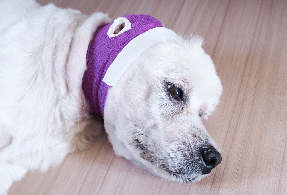
A false cyst is a fluid-filled structure that does not contain a secretory lining. These can form secondary to trauma and hemorrhage. Examples of false cysts include hematomas and seromas. Aural hematomas are common in dogs and are the accumulation of blood within the pinnae of the ear. Aural hematomas are often secondary to ear infections.
Another location that seems to be prone to hematoma development is along the rib cage following trauma. Seromas are similar and can be seen following surgery. Seromas form as serum, the portion of blood not containing red blood cells, and accumulates to fill space between tissue layers.
False cysts should always be checked out by the vet as they require treatment and monitoring in the majority of cases.
4. Sebaceous Cyst
Sebaceous cysts are associated with sebaceous glands and can often be confused with follicular cysts. They are usually a single white or blue looking bump on the skin, and may ooze a thick white or brown cheesy looking discharge, if they burst. Sebaceous cysts usually develop on the head, neck, chest, or legs, and generally do not cause too much concern, but should always be monitored for growth or change in color or appearance.
5. Dermoid Cyst
Dermoid cysts are congenital but may not be appreciated until 9–12 months of age. Dermoid cysts contain a collection of different tissues and are formed in utero prior to birth. The development of the cyst occurs due to the incorrect closure of the skin during development. Cysts may contain bone, cartilage, hair, sweat glands, and other components of skin.
There is also something called dermoid sinus, which is in relation to the spinal cord and is more common in Rhodesian Ridgebacks.
Signs of a Cyst
- Fluid-filled vesicle
- Small bump on or just beneath the skin surface
- Area of hair loss
- Scabby area of skin
- Red, inflamed skin
- Firm, white or blue-colored lump
- Visible discharge coming from the lump or around it
- Discomfort
Diagnosing a Cyst
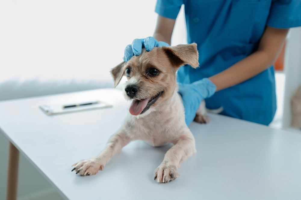
Any skin growth or a cyst cannot be identified based on appearance alone. A fine-needle aspiration performed by a veterinarian is typically the first step in identifying any potential lumps. A needle attached to a syringe is gently inserted into the lump, and the plunger on the syringe is pulled back repeatedly to pull samples of the tissue into the needle. The cell sample pulled is then squirted onto a slide. The slide is carefully stained and evaluated under the microscope. Cells, fluid, and cystic material sampled during a fine-needle aspirate can help provide a diagnosis. Sometimes a sample needs to be sent to an external laboratory for a more in-depth exam.
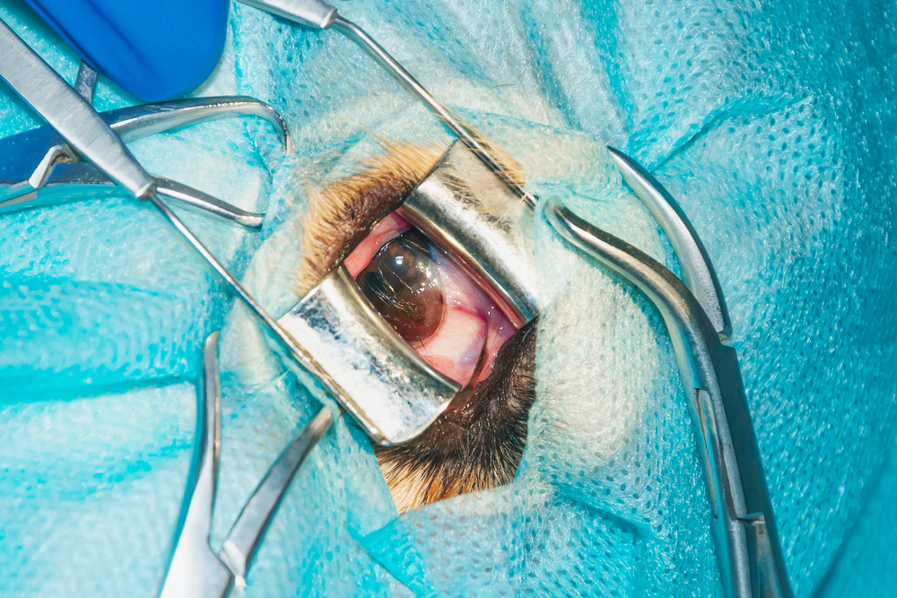
More definitive diagnoses are made through excisional biopsy. In this procedure, the entire cyst is removed under anesthetic and submitted to a laboratory for a histopathologist to evaluate.
How Are Cysts Treated?
Cysts may need to be surgically removed if painful or bothersome for the dog. A cyst that is drained or expressed is likely to refill due to the intact secretory lining that remains. Additionally, manual expression of cysts is not routinely recommended. Manual expression can cause inflammation and discomfort and can lead to infection.
Infected cysts may benefit from topical and oral antibiotics. If a cyst continues to rupture and fails to heal, or is growing in size and causing discomfort to the dog, surgical removal may be warranted. Meanwhile, false cysts should always be checked out by the vet and may require specific treatment depending on their cause and location.
If the lump on your dog is not actually a cyst, but something else, evaluation by your vet and a fine needle aspirate or a biopsy will provide a definitive answer and they will establish an adequate treatment plan, whether it’s monitoring, antibiotic treatment, or surgery.
At Home Care
Cysts should be closely monitored at home for signs of any changes such as growth, redness, discomfort, and infection. A painful cyst may cause a dog to scratch or bite at it. This may lead to damage of the cyst, bleeding, or infection. If your dog is causing trauma to the cyst, an appointment should be made with your veterinarian, alongside attempts to stop the trauma from happening. This can be achieved by covering the cyst or having the dog wear an Elizabethan collar.
In some cases, cysts may not become bothersome for dogs. Routine monitoring is necessary. An appointment should be scheduled with your veterinarian if the cyst ruptures or if signs of infection are present. Signs of infection can include a recent increase in size, discomfort, a red appearance to the skin, or an unusual odor associated with the mass.
If you need to speak with a vet but can't get to one, head over to PangoVet. It's our online service where you can talk to a vet online and get the advice you need for your dog — all at an affordable price!

In Summary
Cysts on the skin are common occurrences impacting our canine companions. Fortunately, most times, cysts are benign. However, it’s best to get them evaluated by your vet as soon as you notice them, as although they may appear to be just cysts, they could also be a sign of underlying skin disease or they may be cancerous lumps. Cysts should be closely monitored for signs of enlargement and infection, and surgical removal of the cyst should be considered if painful, growing, changing in appearance or if infections occur.
Featured Image Credit: Mary Swift, Shutterstock