In this article
View 2 More +Humans have two eyelids per eye—the upper and lower ones. Your dog appears to have two eyelids per eye, but there’s actually a third one that’s hidden from view. So, how many eyelids do dogs have? They have three per eye.
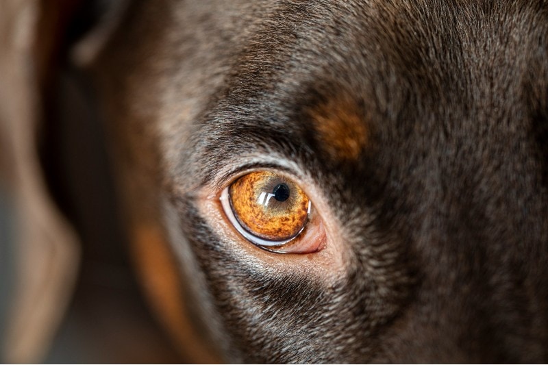
If you’ve ever seen your dog deeply sleeping, you may have noticed a pink triangular membrane in the inner corner peeking through the external eyelids. This is known as the nictitating membrane, or “third eyelid.”

What Is the Third Eyelid?
The third eyelid is found at the inside corner of each eye of domestic dogs, other canines, felines, and other animals. It’s a triangular membrane of conjunctival tissue that covers the surface of the eye to provide protection. The third eyelid also has one of the most important tear glands at its base.
While all breeds have a nictitating membrane, it can vary in its appearance. Some dogs’ third eyelids are very pale or quite dark, but most are pink.
- Protecting the eye from injury
- Keeping the cornea clean and lubricated by spreading tears
- Producing immunoglobulins to protect against infection
- Producing tears


Health Conditions of the Third Eyelid
Although you may not see the third eyelid often, it can develop health conditions that are distinct from those that affect the other eyelids.
Cherry Eye
The most common third eyelid condition is “cherry eye,” or prolapse of the third eyelid gland from its normal position. When this occurs, the eyelid looks like a smooth pink or reddish mass above the edge of the eye. It can happen in one eye or both, simultaneously or at different times.
Cherry eye often becomes obvious when it’s a red, swollen mass that resembles a cherry. It can be large and may cover a portion of the cornea, or it may be small and only visible some of the time.
This can occur when the fibrous attachment that anchors the gland of the third eyelid is weak, enabling the gland to prolapse easily. Several breeds are prone to cherry eye, including Bulldogs, Boston Terriers, Beagles, Lhasa Apsos, Shih Tzus, Cocker Spaniels, and Bloodhounds. It can also occur in brachycephalic breeds of both dogs and cats, or those that have a “squished face” look.
Cartilage Eversion
Cartilage eversion, or scrolled cartilage, is less common than cherry eye and tends to affect large breeds. The third eyelid has T-shaped cartilage inside it, which helps it hold its shape. In young giant breeds, the T area can grow quickly, leading the cartilage to become bent, averted, or scrolled.
When this happens, the third eyelid is “rolled up” and looks like a pink or reddish mass in the corner of the eye. This can look similar to cherry eye, so it may require a thorough examination by a vet to distinguish between the two.
If you want more information or are concerned about the health of your pet, you should contact your vet.
If you need to speak with a vet but can't get to one, head over to PangoVet. It's our online service where you can talk to a vet online and get the advice you need for your pet — all at an affordable price!

How Are These Conditions Treated?
A poorly functioning nictitating membrane and an everted gland leave a dog’s eye at risk of dryness, itchiness, and discomfort. Repeated rubbing and scratching at the membrane can cause other eye injuries, such as corneal ulcers.
With both cherry eye and cartilage aversion, the recommended treatment is surgery. For cherry eye, the gland is returned to its normal position at the base of the third eyelid to ensure that it keeps functioning, while cartilage aversion is treated by dissecting the cartilage excess and removing it. The prognosis is good for both conditions with surgery.


Final Thoughts
Although we may not see them often, dogs have three eyelids that are essential to the health of their eyes. In addition to the upper and lower eyelids that we can see all the time, dogs have a third eyelid that’s hidden in their inner corner. Since some conditions can affect the third eyelid and risk your dog’s eye health, it’s important to pay attention to this eyelid and visit a vet if anything looks strange.
See Also:
- Great Off-Leash Dog Parks in Littleton, CO You Can Visit Today
- There’s a Bump on My Dog’s Eyelid: What to Do
Featured Image Credit: Sabrinasfotos, Pixabay